
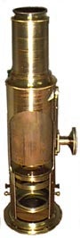
This is a picture of a Drum microscope made in the 1800's. It was built in the midpoint of the development of compound microscopes (containing more than one lens) that began in the very early 1600's (Jansen's 1608 microscope with two lenses) and carries on today. Its predecessor is the single lens, or simple, microscope which came to fame in the mid 1600's due to the work of Antony van Leewenhoek who founded the science of microbiology.
The simple microscope with a single lens
The Leewenhoek microscope was simplicity in itself. It had a single lens mounted on a metal plate with screws to move the specimen across the field of view and to focus its image. The lens was the key and permitted magnification of 70 to 270.A lens works by refraction and is shaped so that the light rays near the center are hardly refracted and those at the periphery are refracted very much more. A parallel beam of light passing through a convex lens is focused to a spot. The distance from the center of the lens to the spot is known as the focal length of the lens. The greater the curvature of a convex lens, the shorter the focal length. The shorter the focal length, the greater the magnification. That is, highly curved lens surfaces are associated with short focal lengths and high magnification. They are called strong lenses.
The magnification M (referred to also as lateral magnification) is defined as the ratio of image and object dimensions.
If an object is magnified a factor of 10 (or times ten) it is usually written x10.
The magnification of a lens can be related to the focal length by using the properties of the human eye which has a nearest distance of distinct vision of about 25 centimeters (10 inches). The magnifying power Mlens of a simple lens viewed by eye is given by the ratio of the apparent size of an object, seen at a distance of 25 cm from the eye to its actual size. If the focal length f of a lens is 2.5 cm (1 inch), then
or a magnification x10.
When using a simple microscope it is useful to have a comparison of distance in view at the same time. A human hair has a diameter of about 70 microns (micrometers = 10-6 meters) where 100 microns = 0.01 centimeters (the symbol for micron is  or m). The view in Fig 1 is of a human hair magnified in an electron microscope.
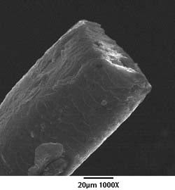
Figure 1. The cut end of E.B. Mayer's hair magnified 1000 times in an electron microscope. The horizontal line indicates a length of 20 microns.
Resolution is the smallest separation between two object points that a given lens (or mirror) can still show as two distinct entities, not one. In practice it is how small are the details you can see with the lens. Resolving power is an instrument property specifying the smallest detail that a microscope can resolve in imaging an ideal specimen. Resolution refers to the detail actually revealed in the image of a real specimen. High resolution refers to small values of the minimum resolvable distance. The resolving powers of optical microscopes are limited by the wavelength of imaging light to about 0.2 microns (200 nanometers). Of course, this limit requires ideal lenses under ideal conditions.
Depth of focus is the distance above and below the geometric image plane within which the image is in focus, Fig. 2.
Figure 2. Depth of focus for a single lens.
As one goes to higher and higher magnification, the depth of field in the sample gets smaller and smaller. It becomes hard to keep the entire specimen in focus. Low-power microscopes have greater depth of focus than do high-power microscopes.
Compound Microscope
The compound microscope, Fig. 3, has two or more lenses. It allows greater magnification, up to 1000x, than the simple microscope by a factor of 4. The objective lens system can be quite complex with doublet lenses (combination of two lenses of different materials) used to correct chromatic aberration which is the spread of an image over a range of colors.
Figure 3. The compound microscope with two lenses.
A beam of light can be brought to a focus due to the refractive index of the lens. The degree of refraction to which light is subjected depends on its wavelength. The refractive index varies with the wavelength of light (a property known as dispersion) and any optical system which depends on refraction will behave differently for different colors. The refractive index of blue light is greater than that of red light. Thus, the focal lengths of simple lenses are shorter for blue light than for red light. In glass lenses (chromatic lenses), chromatic aberration is corrected by the use of combinations of lenses that differ in curvature and dispersion.
With compound microscopes, the image from the eyepiece can be focused on an array of light-sensitive semiconductor devices, known as charge-coupled devices, CCD's. These CCD's convert light into electrons and thus an electronic image is generated which can appear on a TV monitor and can be stored on magnetic or digital media.
Go to Exercise 13
Continue to the Optical vs. Electron Microscopy reading
Go Back to the Previous Page
Page authored by the ACEPT W3 Group
Department of Physics and Astronomy, Arizona State University, Tempe, AZ 85287-1504
Copyright © 1995-2000 Arizona Board of Regents. All rights reserved.