Electron Microscopy
excerpt from Microcosmos by Jeremy Burgess, Michael Marten and Rosemary Taylor
© Copyright 1987, Cambridge University Press, reproduced by permission.

Image Formation in the TEM

The illumination system of the TEM consists of the electron gun and one or, more usually, two condenser lenses. Electrons leaving the heated tungsten filament that forms the electron gun are accelerated towards an anode plate. They pass a biased grid (the Wehnelt cylinder) and travel through a hole in the anode plate before beginning their journey down the microscope column. First they encounter the condenser lenses, which concentrate the electron beam and bring it to a point of focus some way above the specimen plane. The condenser system contains a small physical aperture in the form of a disc of metal such as platinum or molybdenum with a precisely circular hole at its center. This aperture controls both the intensity and the angle of convergence of the electron beam. The operator usually has a choice of three aperture sizes, from a hole 25 micrometers in diameter up to 100 micrometers. Aperture size, together with the precise level of focus of the condenser lenses, form the operator's control of 'brightness' in the final image.
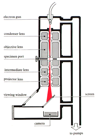

When the electrons encounter the specimen, one of three things can happen. They may pass through it unimpeded. They may be scattered without loss of energy (elastic scattering). Or they may be inelastically scattered; this involves an exchange of energy between the electron beam and the specimen, and may cause the emission from the specimen of secondary electrons or X-rays. Image contrast in the TEM depends on preventing the inelastically scattered electrons from contributing to the image. Their exclusion is accomplished by a second small aperture just below the specimen. This is the objective aperture and, again, the operator usually has a choice of three aperture sizes, which comprise the microscope's contrast control. (In practice, the major influence on image contrast is specimen preparation, as will be seen later.)

The specimen is surrounded by the objective lens (not to be confused with the objective aperture), which magnifies the image of the specimen to a small degree, of the order of x 50. Changes to the current flowing through the objective lens constitute the microscope's 'focus' control. The magnified image from the objective lens is further enlarged by two other lenses below the specimen. They are called the intermediate lens and the projector lens. Both the absolute and relative excitations of these lenses are controlled electronically by a simple 'magnification' control on the microscope's operating console. Most modern TEMs can be set to give a final magnification anywhere in the range x1,000 - x500,000.

Finally, electrons leave the projector lens and strike a screen coated with fluorescent material. The operator sees a continuous image of the specimen, usually green in color, through a viewing window. Fine adjustments to the image are made while viewing it with an externally mounted, low-power binocular microscope.

The control which the operator has over the image is considerable. Brightness, focus, magnification and to some extent contrast are all adjustable. In addition, the operator can improve image quality by careful adjustments of astigmatism correction controls. However, most of the characteristics of the image are predetermined by techniques used to prepare the specimen. A good operator can get the best out of a specimen, but it needs a well-prepared specimen to produce an excellent image.

Go back to the top of this page.

Go on to the next section, Image Formation in the SEM.


Page authored by Paul Perkes and the ACEPT W3 Group
Department of Physics and Astronomy, Arizona State University, Tempe, AZ 85287-1504
Copyright © 1995-2000 Arizona Board of Regents. All rights reserved.