
Introduction
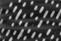
This image shows an atomic force microscope (AFM) view of the data surface of a Compact Disc. AFM is one of the two general types of scanning probe microscopes. The other type is the scanning tunneling microscope (STM). Both types of microscopes show the topography or shape of objects at the nanoscale (one-billionth of a meter). For details, see the Reading on Scanning Probe Microscopy.The Home Activity for this week is to use image processing software to analyze a scanning probe microscope image. After you download the image and software, this exercise can be performed "off-line" (i.e. without using the Web). You will probably want to make a print-out of this instruction page.
Downloading Procedure
Gather the following materials: (Expect to spend at least 20 minutes downloading the image and software if you are using a 28.8 modem; longer for slower modems or busy download times).- Download the image file: disk.tiff. If an image-viewing application starts up (such as JPEGView), please quit it and make sure the CD image is saved to your desktop or somewhere accessible.
- Download image processing software and install it on your computer:
-
a) If you are running Mac OS, you will use NIH Image.
- Go to the NIH Image Web site
- On the Downloads page, choose nih-image161_fat.hqx
- Go there now
- Go to the Scion Corp. Web site and choose Download Scion Imaging Software.
- Scion Corporation distributes this software for free, but they have a survey in progress to find out who is using it. You must fill in the required fields and submit the form before you can download Scion Image PC for Windows 95.
b) If you are running Windows 95, you will use Image PC.
- Go to the NIH Image Web site
- After obtaining the image file and software, quit your browser application (e.g. Netscape or Internet Explorer).
Image Processing Instructions
- Start your image processing software
- Choose File/Open... and choose the file: disk.tiff
This is an Atomic Force Microscope image of a Compact Disc, prior to application of its final protective coating. Each pixel (picture element) in the image represents a measurement of the height of the CD surface for that location. Both music and data CD-ROMs exhibit topography like this at the nanoscale.
- Place your cursor at several locations on the image. Examine the values
displayed in the Info window for each location. (Consider the first number displayed for
each item, not the numbers in parentheses)
- Describe what X, Y, and Value in the Info window represent in this
image.
- Display the data in different colors by choosing Options/Color Tables.
- Use the Profile Plot tool
 to drag a
line along one of the diagonal "lanes" of pits on the CD. (Windows 95 users may need
to move the image aside to see the Plot window).
to drag a
line along one of the diagonal "lanes" of pits on the CD. (Windows 95 users may need
to move the image aside to see the Plot window).The profile plot tool creates a graph showing the value of every pixel it crosses. This is analogous to cutting through the image and looking at it from the side. For this image, the vertical scale (nanometers, nm) is exaggerated by a factor of one thousand compared to the horizontal scale (micrometers,
 m, or
microns).
m, or
microns). - Create a 3-dimensional view of the data:
- Use the rectangular selection tool
 to
select a portion of the CD image.
to
select a portion of the CD image.
- Choose Analyze/Surface Plot
- Use the rectangular selection tool
- File/Close... your surface plot (don't save changes) and create a few more plots.
Experiment with different settings for window size and wireframe/grayscale options.
- On the original image, measure the length and width of some of the pits in the CD:
- Choose the straight line selection tool
 in
the Tools window. On the image, drag a line from the beginning to the end of
the feature you wish to measure.
in
the Tools window. On the image, drag a line from the beginning to the end of
the feature you wish to measure.
- Choose Analyze/Measure.
- The result (in micrometers) will be displayed following Length: in the Info window.
- Choose the straight line selection tool
- Explore some of the other options available in the image processing software. If you "mess up" the image, choose File/Revert to Saved or simply close and reopen the image.
Questions
- How long is the longest pit in the image? ...the shortest?
- How wide are the pits?
- From this image, how deep are the pits?
- A human hair is approximately 50 microns in diameter. How many "lanes" of CD pits would fit across this distance?
- CD data are read at a constant linear velocity of 1.3 meters per second. To facilitate this, does a CD player need to spin faster when reading the inner portion or the outer portion of a CD? Explain your answer.
To learn more about CDs and other information storage mediums: Click here
To learn more about ASU's Interactive Nano-Visualization for Science and Engineering Education: Click here
To learn more about the Image Processing for Teaching project at University of Arizona: Click here
Go to the Scanning Probe Microscopy Reading
Activity by LuAnn Dahlman, IPT project with support from IN-VSEE, Arizona State University
Copyright © 1995-2000 Arizona Board of Regents. All rights reserved.
Page authored by the ACEPT W3 Group
Department of Physics and Astronomy, Arizona State University, Tempe, AZ 85287-1504
and LuAnn Dahlman, IPT project