HEAD, NECK And SHOULDER ASSESSMENT
Assessment of the head is important as it reflects an evaluation of the central nervous system. Often assessment of the newborn's head reveals minor variations from normal which are not usually of clinical significance. Because parents often focus on the head first, it is important to reassure them of the normal variations.
Molding
Assess the appearance, shape and presence of molding.
|
|
|
|
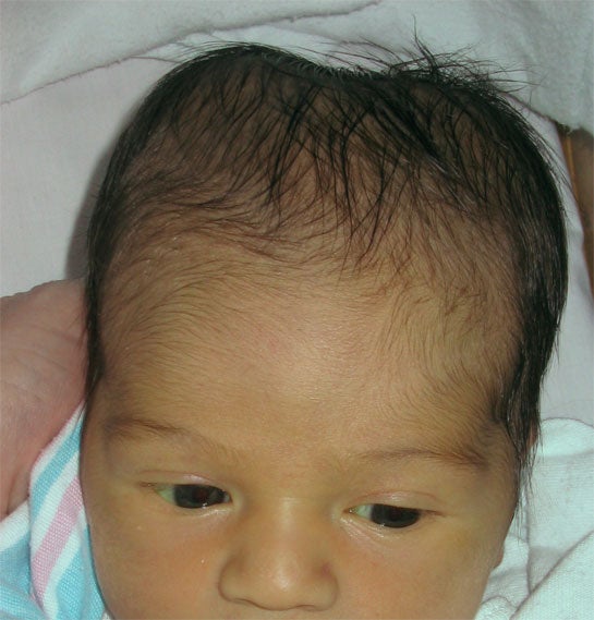
Molding at Birth (Elsevier) |
Overriding Sutures and Molding (Dr. Janelle Aby) |
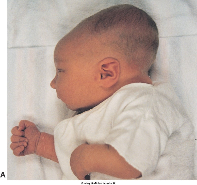
The left picture (above) shows how the head is elongated in molding. The right picture shows the angled indentation of the sutures where the bones overlap each other. This is called overriding sutures.
|
|
|
|
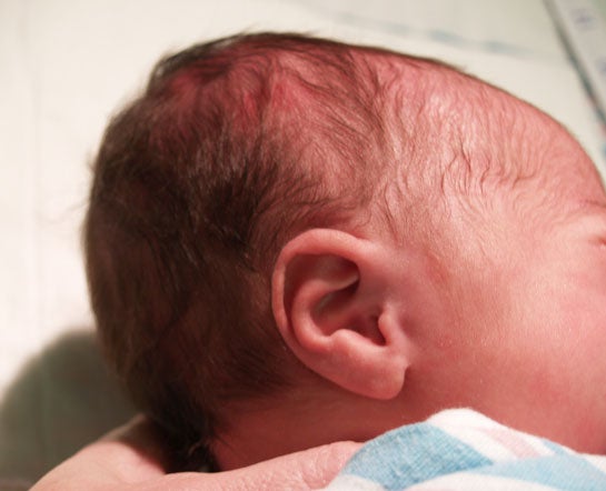
Cranium Molding during Vaginal Birth |
Molding After Birth and at 24 hours |
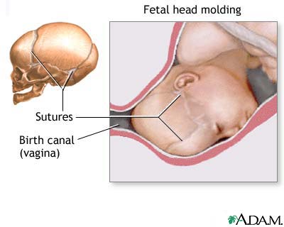
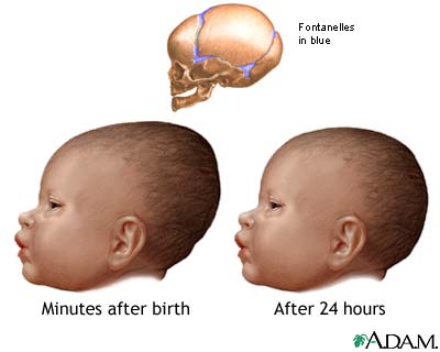
The left picture shows how the sutures allow the bones of the cranium to mold or compress to facilitate movement move through the vagina. The right picture shows the infant cranium on the first day of life and 24 hours later. Infants born via c-section birth or breech birth do not have molding. The temporary cranial asymmetry returns to a normal shape by the end of the first week.
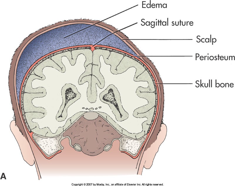
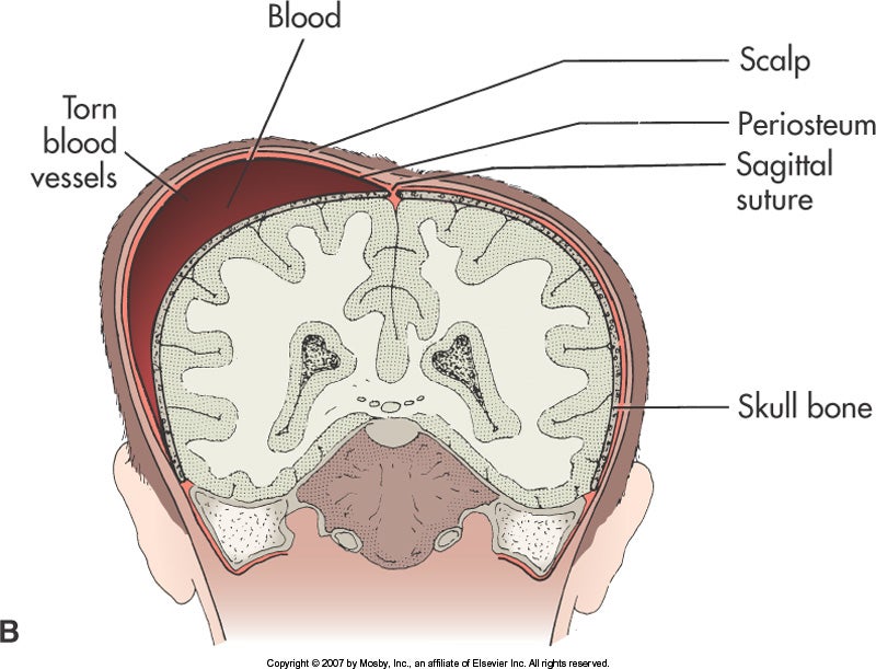
Caput succedaneum or Cephalohematoma
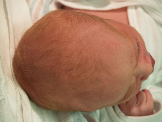
With the infant lying on his back, cradle the infant's head in your hands. Feel for softness or "squishiness" on one side or both sides of the head. This can either be a "caput"(A) or a cephalohematoma (B). The swelling with caput disappears by the end of the first week whereas the swelling of cephalohematoma may take six weeks. Caput can cross over suture lines whereas cephalophematoma may be confined to one cranial bone. Neither of these variations are present with a c-section birth.
|
|
|
|
Caput Succedaneum |
Cephalohematoma (Elsevier) |
Pictures used with permission Dr. Janelle Aby, Stanford Medical School, Palo Alto, CA
The pictures above show an infant head with a caput. The first picture shows molding of the head, caput and swelling at birth. The second picture shows pitting and bruising on the head. The third picture shows an almost normally shaped head at 24 hours after birth.
Pictures used with permission Dr. Janelle Aby, Stanford Medical School, Palo Alto, CA
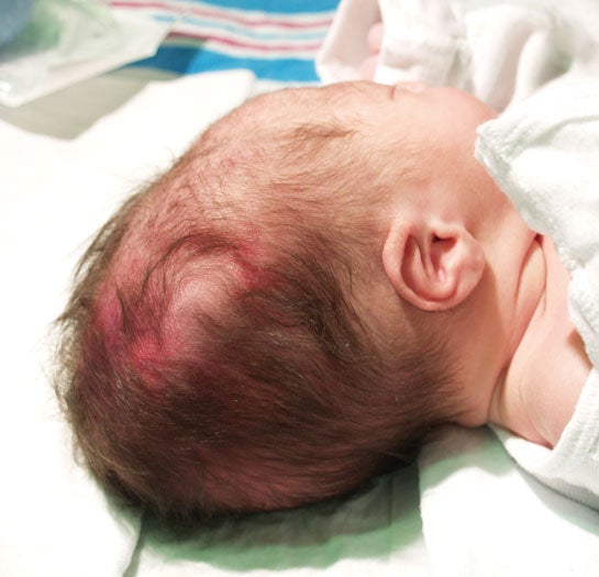
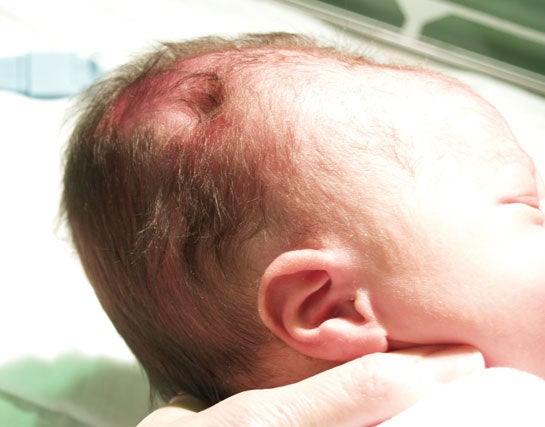
These pictures show a cephalohematoma. The infant has swelling over the right parietal bone (left). The right picture shows swelling over the left parietal bone.
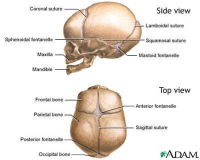
Anterior and Posterior Fontanels
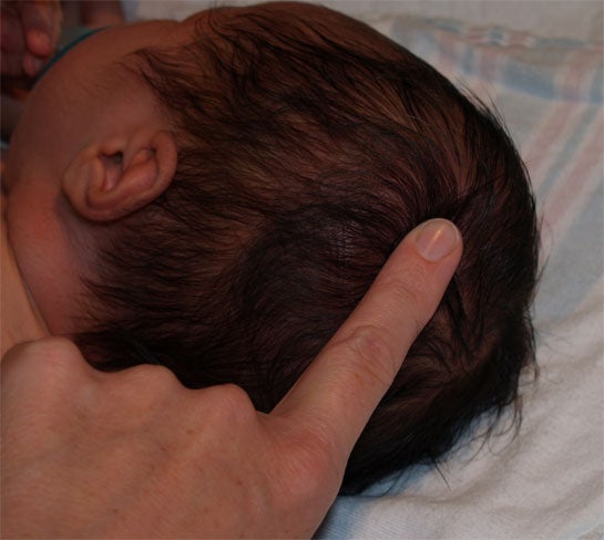
While holding the head with one hand, move the "pointer finger" of the other hand to the front of the head to palpate the triangular shape of the anterior fontanel. Measure the length and the width. A rough estimate can be obtained by using the width of the finger as 1 cm and measuring the length and width in this manner. If a more exact measurement is needed, use a tape measure.
From the anterior fontanel trace your finger down the juncture of the sagittal suture and the lambdoidal suture to the posterior fontanel. This may be barely palpable. The posterior fontanel disappears at 6 weeks, whereas the anterior fontanel disappears at about 18 months. Although the anterior fontanel is described as a 5cm diamond shape, the fontanel size varies with the degree of molding. It may be difficult to palpate the fontanels if there is a great deal of molding. As the molding resolves, the fontanel size will increase. Depressed fontanels can be due to dehydration whereas bulging fontanels can be due to hemorrhage, infection or tumor. Large flat and soft fontanels could be due to hydrocephaly, hypothyroidism or malnutrition.
Neck and Shoulders
Observe the neck. Check the movement and position of the head. Restricted movement or holding the head at an angle could be due to torticollis (wryneck) or opisthotonos. Absense of head control may be due to prematurity or Down's Syndrome.
Use fingers from both hands to palpate simultaneously for intactness and crepitus of the clavicles. Assess range of motion of arms. One arm may also have decreased range of motion with clavicular fracture. This should be assessed with any difficult birth, particularly with shoulder dystocia.
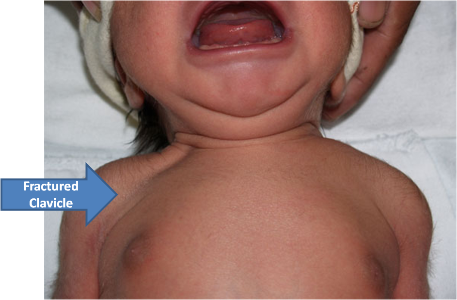
This unhappy newborn has a right fractured clavicle probably due to a difficult birth with shoulder dystocia.
Picture used with permission Dr. Janelle Aby, Stanford Medical School, Palo Alto, CA
